Ultrasound

Ultrasound is an amazing tool in the antenatal care and can be used from the first strokes of the fetal heart around week 6, until the baby is born. Ultrasound gives us the opportunity to have a look inside the uterus to gather information regarding the pregnancy and by that provide the best care for mother and baby.

What information can we get from ultrasound?

  • Prior week 11 :

    We can see how many fetuses there are, confirm that the pregnancy is well established inside the uterus and also measure the fetus to find the approximate length of the pregnancy

  • Week 11-14

    Now the anatomi is almost fully developed and makes this a perfekt time to conform the development . This is also when you want to have a look at the neck/NT. Measuring the fetus will give good information regarding the length of the pregnancy. You can usually confirm the gender from week 13+0.

  • Week 18-20

    In Norway this is when you are given the final due date. You also check the anatomy and determine the position of the placenta. This exam is usually done in the hospital, SUS and is part of the free public health services. You can choose to have this exam with us.

Measuring growth of the fetus

Can be done any time after the 24th week of pregnancy. When calculating fetal weight, you use the due date as a benchmark to determine whether the fetus weighs more or less than average. In order to comment on how the fetus is growing it is necessary to take several measurements over time.

If there is any reason to suspect any illness or defect, you will be referred to the hospital.

Is ultrasound dangerous?

There is no documentation indicating that properly administered ultrasound examinations are dangerous.

Ultrasound examinations have been used during pregnancies since the 1960’s. Nonetheless, it is the responsibility of the sonographer to ensure that the unborn child not be exposed to unnecessary risk, in connection with the use of high frequency energy.

2D Ultralyd

I can give you 2-D ultrasound examination at any time during the pregnancy. Each time, you will receive a complete fetal examination.

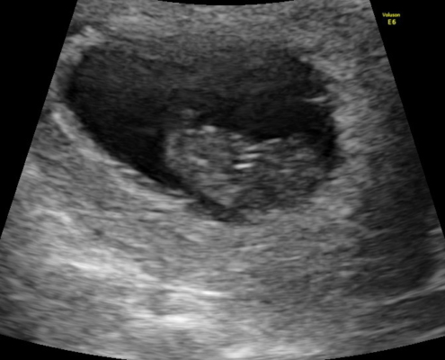
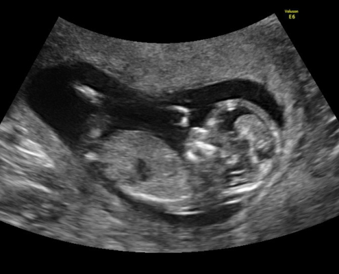
The photos visible on the screen are 2-D cross sections of the uterus and fetus. This examination provides detailed information about the anatomy of the fetus.

3D/4D Ultrasound

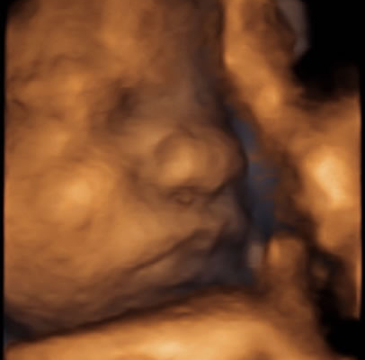
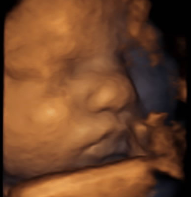
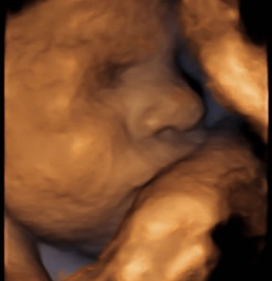
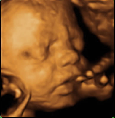
3-D & 4-D ultrasound is developed, based on 2-D ultrasound pictures, taken in several directions.

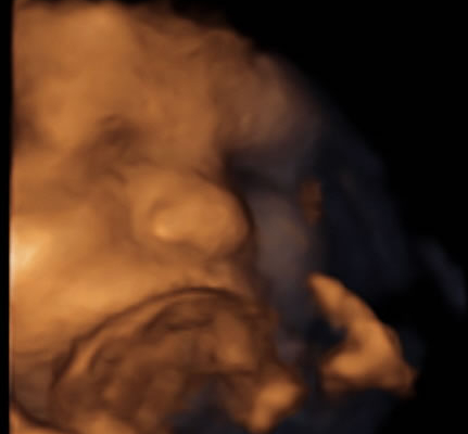
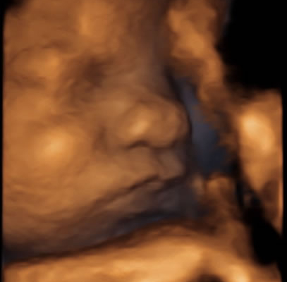
3-D ultrasound allows one to see length, breadth and height in one picture; but it does not show any movement.

4-D ultrasound shows the fetus in movement, using 3-dimensional photographs. The fourth dimension represents time.

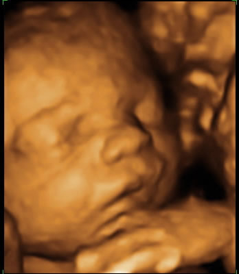
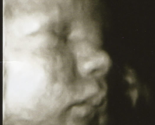
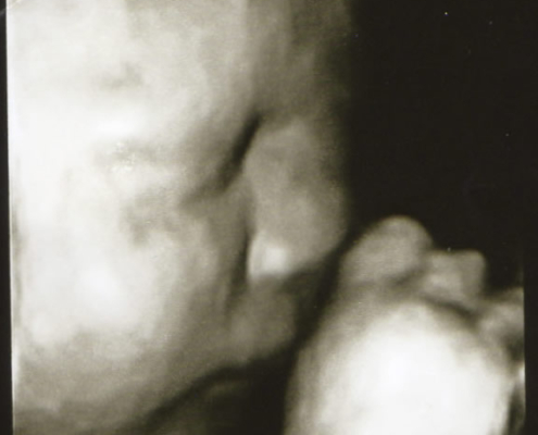
3D/4D can be used through the whole pregnancy and is a good way of showing the fetus. For close-up pictures of the face it`s recommended around week 28-32.

In this exam I will also do an estimation of the growth/est weight of the fetus, as well as cheque the fetal water and position.

In order to obtain good 3-D and 4-D pictures or film, it is important that:

  • the fetus is surrounded by amniotic fluids in the area that is being photographed, as fluids provide a “window” to the fetus.
  • the fetus is lying in a position which is accessible. For example, that means
  • that the fetus cannot have its face pushed against the wall of the uterus or behind the mother’s spine.
  • the mother does not have a layer of fat below the surface of her skin, because fat disperses sound waves, and then the ultrasound pictures become fuzzy.